Summary: Should PSMA PET CT Be Used Earlier in Diagnosing Prostate Cancer?
PSMA PET CT and MRI scans offer highly accurate prostate cancer detection, but their role in early diagnosis is still under research.
- What is PSMA PET CT? An imaging test using a radioactive tracer to detect prostate cancer.
- Current Use: Primarily for high-risk or recurrent cases.
- Benefits: Detects small lesions, improves treatment planning, and reduces unnecessary biopsies.
- Early Diagnosis Potential: Promising but requires more research.
Prostate cancer is the second-most common cancer among men, with roughly 1 in 8 men receiving a prostate cancer diagnosis in their lifetimes.
Over time, diagnostic options for prostate cancer tend to increase and improve. As newer but proven tests for prostate cancer become established, it’s only natural to wonder if they could be used earlier in the diagnostic process.
PSMA PET CT and MRI scans have been shown to better detect the spread of prostate cancer in the body than many standard imaging approaches, as the National Cancer Institute explains.
So, what are PSMA PET CT and MRI scans? And should this scan be used earlier for primary diagnosis of prostate cancer?
Keep reading to learn more about PSMA PET scans and current research related to their use in earlier diagnosis of prostate cancer.
What Are PSMA PET CT and MRI Scans?
Defining PSMA PET CT and MRI Scans
A PSMA PET scan is an imaging test that uses a radioactive tracer substance to identify the presence of prostate cancer cells. Let’s break down this term to better understand its two key components.
PSMA stands for prostate specific membrane antigen. As the Cleveland Clinic explains, PSMA is a protein found on the exterior of prostate cells. Research published in the peer-reviewed journal Reviews in Urology elaborates that all prostate tissue expresses PSMA, with cancerous tissue showing increased expression of PSMA.
Generally, as prostate cancer increases in severity, more PSMA is expressed by prostate tissue.
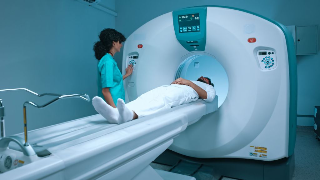
A positron emission tomography (PET) scan is a type of medical imaging distinguished by its use of a radioactive tracer injected into a patient. This radioactive tracer helps medical professionals visualize a variety of changes in the body’s cells, including but not limited to identifying the presence of cancerous cells.
The amount of radioactivity added to the body by the radiotracer is small. It has a low risk of causing side effects, as the Mayo Clinic explains.
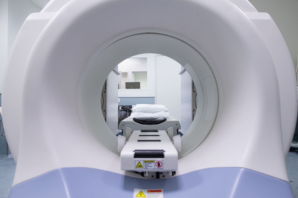
CT scans (formerly known as CAT scans) and MRIs are both types of medical imaging. They allow for a more detailed view of the interior of the body. Both MRI and CT scans can be, and often are, combined with a PET scan to give medical professionals more visibility and context when interpreting the results of these imaging tests.
So, a PSMA PET CT scan is a combination of a PSMA PET scan and CT scan of the prostate gland and other parts of the body. A PSMA PET MRI scan combines the PSMA PET scan with an MRI of the prostate gland and other parts of the body.
Current Diagnostic Use of PSMA PET CT and MRI Scans
PSMA PET CT and MRI scans are currently used for patients in specific circumstances – they are not a common procedure for all patients that are at risk of or diagnosed with prostate cancer.
Healthline points out that PSMA PET scans are often ordered by a physician when a patient both:
- Has a prostate cancer diagnosis, and
- Displays risk factors for the spread of cancer cells to other parts of the body (metastasis).
PSMA PET CT and MRI scans are also used when patients have been treated for prostate cancer but still have detectable levels of the disease. Similarly, these tests are also used after a patient is treated for prostate cancer, but follow-up testing, such as PSA tests, show a potential return of cancer.
The role of PSMA PET CT and MRI tests for prostate cancer early detection is still being explored.

Why are PSMA PET Scans Important for Prostate Cancer Patients?
PSMA PET scans are important for prostate cancer patients because they can provide better visualization of cancerous cells within the body as compared to other types of medical imaging. By combining PSMA PET scans with MRIs and CT scans, medical professionals gain additional and valuable context about the state of prostate cancer in the body.
PSMA PET scans have also been shown to detect small lesions in the body, meaning these scans can detect prostate cancer spread earlier than other tests.
A study of 30 patients with recurrent prostate cancer and suspected metastases in their lymph nodes was published in the peer-reviewed journal Theranostics. PSMA PET scans on the patients detected small lesions with high accuracy, above 95% in terms of positive predictive value, close to 90% negative predictive value, and about 95% in terms of overall accuracy.
With their accuracy in terms of small lesions, these scans may help clinicians better answer the question of how to detect prostate cancer spread early on.
A study published in the peer-reviewed Journal of the American Medical Association found that, for simulated patients with recurrent prostate cancer, upfront PSMA PET scans could lead to 75 fewer deaths and 988 more life-years per every 1,000 patients in comparison with traditional imaging.
A review published in the peer-reviewed Japanese Journal of Radiology stated the following about PSMA PET scans in relation to prostate cancer: “This modality offers high detection capacity for prostate cancer lesions and is anticipated to significantly impact the treatment of prostate cancer with application in various scenarios of prostate cancer treatment.”
Additionally, PSMA PET scans may help to reduce the need for unnecessary biopsies and treatments due to the scan’s strong detection capabilities. With clearer insight into the presence and spread of prostate cancer, more efficient treatment and fewer unnecessary procedures are possible.
Finally, the way in which PSMA PET scans function makes them the superior tool for detecting the spread of prostate cancer to lymph nodes and bones. That enhanced detection can improve treatment plans for patients. For example, if a PSMA PET/CT shows a single small lymph node being positive for cancer spread, that lymph node can be hit with targeted and concentrated radiation instead of just blasting the entire pelvis.
Should PSMA PET Scans Be Used Earlier in Diagnosing Prostate Cancer?
Prostate cancer has few distinct symptoms, especially in its early stages. A simple answer to the question of “How to detect early signs of prostate cancer?” is not yet clear. However, improvements in detection and diagnosis may help more patients and their physicians identify prostate cancer earlier on, before it develops and spreads in the body.
So, should PSMA PET CT scans and MRI scans be used earlier in diagnosing prostate cancer?
Research that could answer this question is ongoing. A 2022 critical review published in the peer-reviewed journal Cancers identified a wide range of areas that need more attention in terms of evaluating PSMA PET scans for use in the primary diagnosis of prostate cancer.
Practical and diagnostic factors both play a role in the value of PSMA PET scans for general early detection of prostate cancer, i.e. for patients who do not present metastatic risk factors or have not already completed prostate cancer treatment.
As with any other diagnostic procedure, cost and accessibility are both important factors. A primary diagnostic procedure can only be beneficial on the broad scale for patients showing a risk of prostate cancer if the procedure is both accessible and affordable.
Similarly, while PSMA PET scans have a high degree of accuracy in identifying lesions in specific subgroups of patients, more research is needed into their effectiveness for use in the much larger group of patients showing potential signs of prostate cancer.
A 2024 overview of the same topic, published in the peer-reviewed journal Seminars in Nuclear Medicine, identified benefits related to diagnosis.
These include the sensitivity and specificity PSMA PET scans provide for identifying metastases in lymph nodes and bones. The value these scans offer in terms of secondary biopsies for patients with negative results from primary biopsies and negative multiparametric MRI (mpMRI).
Similarly, PSMA PET scans could help to guide biopsies for patients where this scan is positive but MRI results are negative. By targeting areas of concern revealed by the PSMA PET scan, false negative results from a biopsy would likely be less common.
The overview states that the superior diagnostic accuracy offered by PSMA PET scans for primary staging affects roughly 25% of patients. However, it also notes that studies are needed to determine how using these scans will influence patient outcomes.
Early use of these scans may benefit high-risk patients, but routine early screening for most patients is still under debate. For patients with intermediate-risk prostate cancer, especially those with early spread, PSMA PET scans could be helpful as well.
PSMA PET CT and MRI scans show plenty of promise in terms of earlier use and more widespread use for initially diagnosing prostate cancer.
More research is needed to determine exactly when and under what circumstances these scans should be used as a frontline, early, and/or widespread screening procedure.
Treatment Options for Prostate Cancer
Prostate Laser Center is proud to provide minimally invasive, MRI-guided treatment for prostate cancer. Request a free consultation by completing the simple form below.
Learn more about the use of MRI, an established and proven tool, for prostate cancer diagnosis – get the facts today!
NOTE: The information provided on this website is general medical information and does not establish a physician-patient relationship. Please discuss your particular situation with a qualified medical professional.
